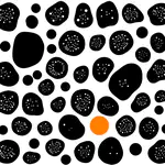
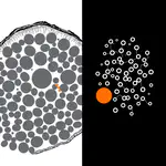
Single-cell sequencing tells you what a cell is, but to read it, it dissolves the tissue — and throws away every cell’s address. Imaging-based spatial transcriptomics keeps the map: it images individual RNA transcripts inside intact tissue at subcellular resolution. The catch is that it turns biology into a hard microscopy problem — you have to segment each cell and assign every molecule to the right one — and then learn what the arrangement means. Tools like Baysor solve the segmentation; foundation models like Nicheformer learn the niche. It’s the imaging×omics intersection at the heart of the lab, and the tissue-context layer a virtual cell is still missing.